
FIGURE 4.
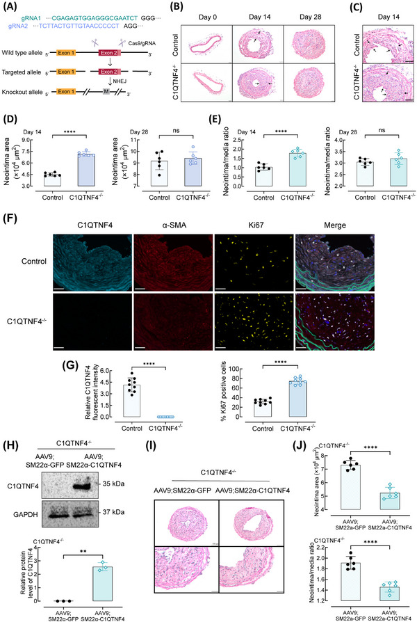
C1QTNF4‐knockout exacerbates wire injury–induced neointimal thickening in mouse carotid arteries and restored C1QTNF4 expression in VSMC exerts a rescue effect. (A) Schematic of the genome engineering targeting strategy of the CRISPR/Cas9‐mediated C1QTNF4 conventional knockout (C1QTNF4−/−) mouse model. Exon 2 was selected as target site. NHEJ, non‐homologous end joining. M, mutation. (B, C) Representative HE staining of 8‐week‐old C1QTNF4−/− and sex‐matched littermate control mouse common carotid arteries on the 0th, 14th and 28th days after wire injury. Scale bar, 50 μm. (D, E) Quantitative analysis of the neointimal area on the 14th and 28th days (D), and the neointima/media ratio on the 14th and 28th days (E) (± SEM; n = 6 per group). (F) Representative immunofluorescent staining of control and C1QTNF4−/− mouse common carotid arteries at 14 days after wire injury. Slides were simultaneously stained with a multiplex QIF panel containing C1QTNF4 (cyan), α‐SMA (red), Ki67 (yellow), EL (green) and DAPI (blue). Scale bar, 30 μm. (G) Quantification of the C1QTNF4 expression and Ki67 positive cells in the injured arteries (± SEM, n = 9). (H) 6‐week‐old C1QTNF4−/− mouse infected with AAV9; SM22α‐C1QTNF4 vectors and sex‐matched C1QTNF4−/− mouse infected with AAV9; SM22α‐GFP vectors. Western blot of C1QTNF4 in the common carotid arteries VSMCs of C1QTNF4−/−; AAV9; SM22α‐GFP mice and C1QTNF4−/−; AAV9; SM22α‐C1QTNF4 mice. Protein levels of C1QTNF4 were assessed by Western blot analysis (± SEM, n = 3). (I) Representative HE staining of C1QTNF4−/−; AAV9; SM22α‐C1QTNF4 and C1QTNF4−/−; AAV9; SM22α‐GFP mouse common carotid arteries on the 14th day after wire injury. Scale bar, 100 μm, 50 μm. (J) Quantitative analysis of the neointimal area and neointima/media ratio on the 14th day. ± SEM, n = 6 per group. **p < .01, ****p < .0001; NS indicates no significance.