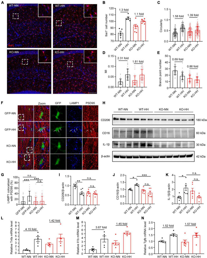
FIGURE 4.
CX3CR1 deficiency attenuates HH-induced-M1-type microglia and phagocytosis of synapses by microglia. (A) Immunofluorescence labeling of microglia/macrophage marker Iba1 in the hippocampal CA1 region, Scale bar = 100 μm. (B–E) Statistics of panel (A), including the number of Iba1+ cells [(B), n = 5], circularity [(C), n = 111–130, brain sections from 5 mice of each group were stained, and 20–30 cells of each mouse were quantified], MI [(D), n = 33 to 43, brain sections from 5 mice of each group were stained, and 6 to 7 cells from CA1 regions of each mouse were quantified], and branch numbers [(E), n = 45, brain sections from 5 mice of each group were stained, and 9 cells of each mouse were quantified]. (F) Immunofluorescence labeling of LAMP1 and PSD95 in the hippocampal CA1 region of CX3CR1-GFP and KO mouse, Scale bar = 10 μm. (G) Co-localization ratio of LAMP1 to PSD95 in GFP+ cells in panel (F) were determined. n = 35–41, brain sections from 6 mice of each group were stained, and 5–7 cells of each mouse were quantified. (H–K) Western blot detection of CD206, CD16, and IL-1β levels in mouse hippocampal tissue, n = 6. qRT-PCR was performed to detect the changes in the levels of inflammatory factors Tnfa (L), Il1b (M), and Tgfb (N) in the hippocampus, n = 5. *p < 0.05, **p < 0.01, ***p < 0.001, and n.s. indicates no statistical difference.

