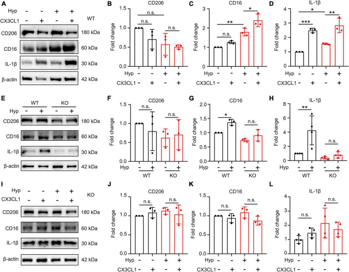
FIGURE 5.
CX3CL1/CX3CR1 signal mediates hypoxia-induced-M1 type microglia. Primary microglia were treated with 1% O2 for 20 h followed by the co-treatment with 50 ng/ml CX3CL1 for 4 h. (A–D) Western blot detection of CD16, CD206, and IL-1β levels in microglia after hypoxia and CX3CL1 treatment (A), and the grayscale values of each band were counted (B–D), n = 3. (E–H) Primary microglia generated from wild-type (WT) or CX3CR1-deficient (KO) mice were treated with 1% O2 for 24 h. The cells were lysed to determine CD16, CD206, and IL-1β levels by Western blot, n = 3. (I–L) Primary KO microglia were treated with 1% O2 for 20 h followed by the co-treatment of 50 ng/ml CX3CL1 for 4 h. Cells were lysed to detect CD16, CD206, and IL-1β levels by Western blot, n = 3. *p < 0.05, **p < 0.01, ***p < 0.001, and n.s. indicates no statistical difference.

