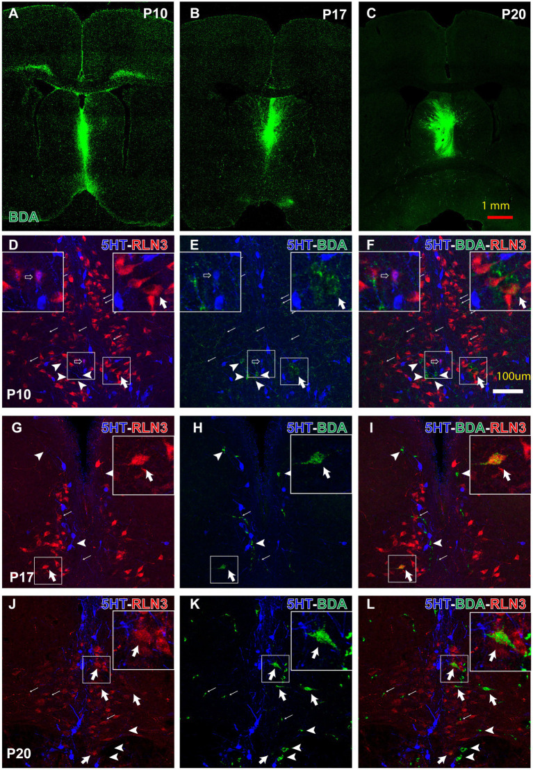
Figure 9.
Anterograde and retrograde labeling in the pontine tegmental raphe/NI border at key postnatal ages, 3 days after prior BDA-3kD injections into the MS, combined with RLN3 and 5HT immunohistochemistry. (A–C) representative injection sites at P10 (survival P10–P13) (A), P17 (survival P17–P20) and P20 (survival P20–P23) Calibration bar, 1 mm. (D–F) appearance of triple 5HT, RLN3 and BDA labeling in the NI between P10–P17. Inset, (left) is a magnified view of the boxed area (left), (illustrating a 5HT + RLN3 double-labeled neuron); inset (right) is a magnified view of the boxed area (right), illustrating a RLN3 + BDA double-labeled neuron. (G–I) appearance of 5HT + RLN3 + BDA triple-labeling in the NI between P17–P20. Inset is a magnified view of the boxed area, illustrating a RLN3 + BDA double-labeled neuron. (J–L) 5HT + RLN3 + BDA triple-labeling in coronal sections at the level of the NI after a BDA injection into the MS at P20 and processing at P23. Inset is a magnified view of the boxed area, illustrating a BDA + RLN3 double-labeled neuron. From (D) to (L), arrows indicate anterogradely-labeled BDA fibers, arrowhead indicates single, BDA retrogradely-labeled neurons, open arrow indicates RLN3 + 5HT double + labeled neurons, and thick arrow indicates RLN3 + BDA double-labeled neurons. Calibration bar in (L) for (D–L), 100 μm.

