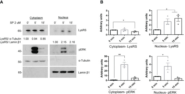
Figure 1.
LysRS translocates to the nucleus after MRGPRX2 activation in mast cells (LAD2). (A) Cells were activated with 2 µM substance P (SP) at various times. Cytoplasmic and nuclear fractions were isolated, and western blots for LysRS and pERK were performed. α-tubulin and Lamin β1 markers for cytoplasm and nuclei, respectively, were used to assess cellular fraction purification and as loading controls. (B) The unpaired t-test was used for statistical analysis (*p < 0.05*, **p < 0.01). Experiments are the mean ± SEM (n=3).

