Figure 4.

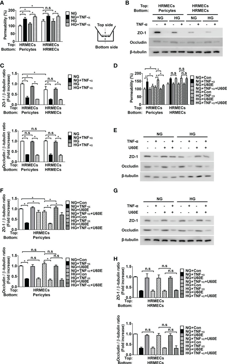
Effect of U60E on the in vitro permeability in co-cultures of pericytes and human retinal microvascular endothelial cells (HRMECs) and the tight junction protein expression in HRMECs. (A) Pericytes and HRMECs incubated on the indicated side of the Transwells as depicted at right. Pericytes and HRMECs were treated with tumor necrosis factor α (TNF-α) (100 ng/ml) under normal glucose (NG; 5 mM glucose) or high glucose (HG, 30 mM glucose) for 72 h. The permeability was measured using Evans blue dye (n = 5). (B) The tight junction protein expression of ZO-1 and occludin was measured from the top side HRMECs lysates obtained by (A). (C) Quantitative densitometric analysis in (B) to calculate the ratio of each protein to β-tubulin. The bar graph represents the means ± standard deviation (SD) (n = 3). (D) Pericytes and HRMECs were incubated on the indicated side of the Transwells and then treated with U60E (20 μg/mL) and/or TNF-α under conditions exposed to NG or HG for 72 h. The permeability was measured using Evans blue dye (n = 5). (E–H) The tight junction protein expression of ZO-1 and occludin was measured from the top side HRMECs lysates under conditions for co-culture of pericytes and HRMECs (E, F) or conditions for culturing only HRMECs (G, H) obtained by (D). Quantitative densitometric analysis was performed to calculate the ratio of each protein to β-tubulin (F, H). The bar graph represents the means ± SD (n = 3). No significance (n.s.) indicates P > 0.05, *P < 0.05.
