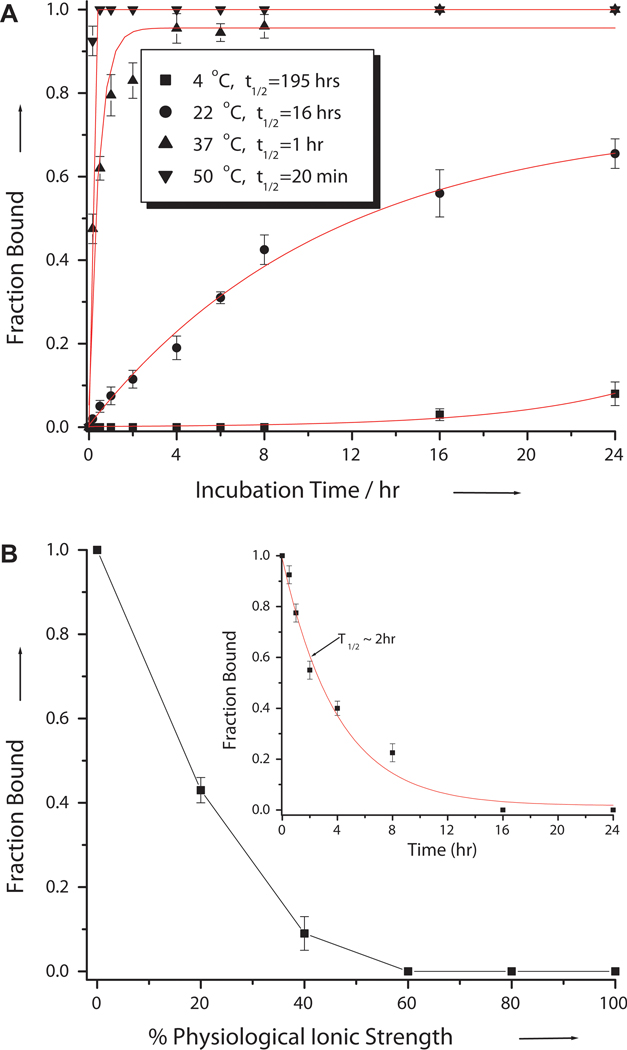
Figure 3.
Effects of (A) temperature and (B) ionic strength on the invasion efficiency. The amounts of fraction bound was determined by gel-shift assays following incubation of 0.1 μM DNA containing PM binding site with 2 μM of R-MPγPNA1 in (A) 10 mM NaPi buffer for 16 hrs at the indicated temperatures, and (B) different percentages of physiological ionic strength (2 mM MgCl2, 150 mM KCl, 10 mM NaPi) at 37 °C for 16 hrs, followed by electrophoretic separation and SYBR-Gold staining. (B) Inset: Profile of the R-MPγPNA1•DNA complex dissociating as a function of time after reconstituting the sample with 100% physiological ionic strength. t1/2 is defined as the time it took to reach 50% binding.

