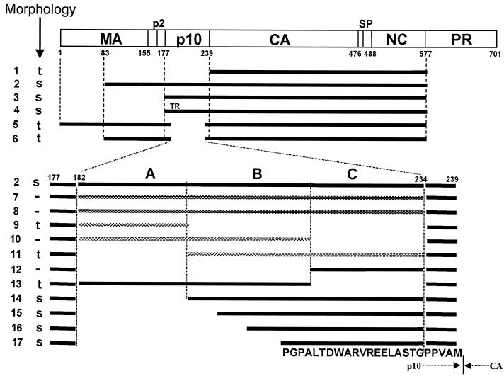
FIG. 1.
Diagrammatic representation of assembly results. The rectangle shows the structure of the Gag protein, with vertical lines representing cleavage sites and numbers representing the number of amino acid residues from the N terminus, using the standard numbering for the Prague C strain of RSV (32). Horizontal bars indicate the structures of the proteins studied for their assembly properties from both in vitro and particle extraction experiments. Black bars represent viral sense sequences. Cross-hatched bars indicate either antisense RSV sequences or sequences from other retroviruses. Regions A, B, and C in p10 are segments of approximately 20 aa residues each. All constructs were placed into the pET3XC vector by common cloning techniques, propagated in E. coli DH5α cells, confirmed by sequencing, and transformed into BL21(DE3)/pLysS cells for protein expression and purification. ΔMBDΔPR, dp10 (called Δp10.52 in reference 25), and CA-NC have been described previously (5, 6, 25). ΔMBDdp10 combines the deletions of ΔMBDΔPR and dp10. DNA segments encoding the C-terminal 62 aa residues of M-MuLV p12 or HIV-1 MA or various segments of p10 (AB, BC, and C) were amplified by PCR from the appropriate viral clones, using primers encoding a SpeI site, and inserted into the unique SpeI site in ΔMBDdp10. Lines: 1, CA-NC; 2, ΔMBDΔPR; 3, p10-CA-NC; 4, p10TR-CA-NC; 5, dp10; 6, ΔMBDdp10; 7, C-terminal 62 aa of MMLV p12; 8, C-terminal 62 aa of HIV-1 MA (BH10 strain); 9 to 11, insertions of antisense sequences derived from DNA coding for p10; 12, segment C; 13, segment AB; 14, segment BC; 15, segment C plus the last 15 aa of segment B; 16, segment C plus the last 10 aa of segment B; 17, segment C plus the last 5 aa of segment B. The proteins shown in lines 7 to 14 contain the inserted dipeptide Thr-Ser between the wild-type Gly and Pro residues five residues from the C terminus of p10. The proteins shown in lines 15 to 17 contain the wild-type sequence at this location. The 25-aa sequence corresponding to the minimal insertion sufficient for spherical particle formation is shown at the bottom, with the boundary between p10 and CA marked. s, formation of spheres; t, formation of tubes; −, no regular assembly; TR, Thr-Arg insertion. Assembly for all of the proteins shown was tested both in vitro with purified protein and in E. coli lysates, with the same results, shown in the column marked “Morphology.”

