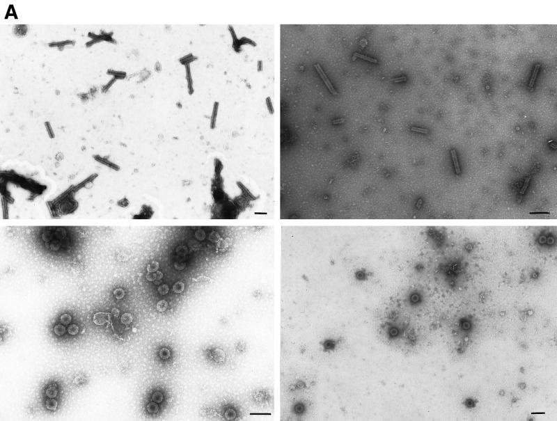
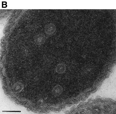
FIG. 2.
Morphology of CA-NC and p10-CA-NC particles assembled in vitro and within E. coli. (A) Electron micrographs of negatively stained particles. Samples were adsorbed onto Formvar- and carbon-coated grids for 2 min and stained with 2% uranyl acetate (pH 5.2) for 20 s. Upper left, CA-NC protein assembled into tubes in vitro; upper right, CA-NC tubes from crude extracts; lower left, p10-CA-NC protein assembled into spheres in vitro; lower right, p10-CA-NC spheres from crude extracts. Bars = 100 nm. (B) Thin-section electron micrograph of VLPs formed within E. coli cells expressing p10-CA-NC protein. Cells were pelleted and fixed for 2 h in 0.1 M sodium maleate (pH 5.2)–3% glutaraldehyde and then washed in 0.1 M sodium cacodylate, pH 7.4. The samples were postfixed for 2 h in 1% OsO4–0.1 M sodium cacodylate (pH 7.4), quickly rinsed in 0.1 M sodium maleate (pH 5.2), and then washed extensively in the same buffer. The samples were then stained with 1% uranyl acetate–0.1 M sodium maleate (pH 6.0), washed in 0.1 M sodium maleate (pH 5.2), and serially dehydrated with 50, 70, 95, and 100% ethanol and 100% propylene oxide. Pellets were embedded in 50% propylene oxide–standard Spurr, and thin sections were stained with 2% uranyl acetate and then Reynold's lead citrate.


