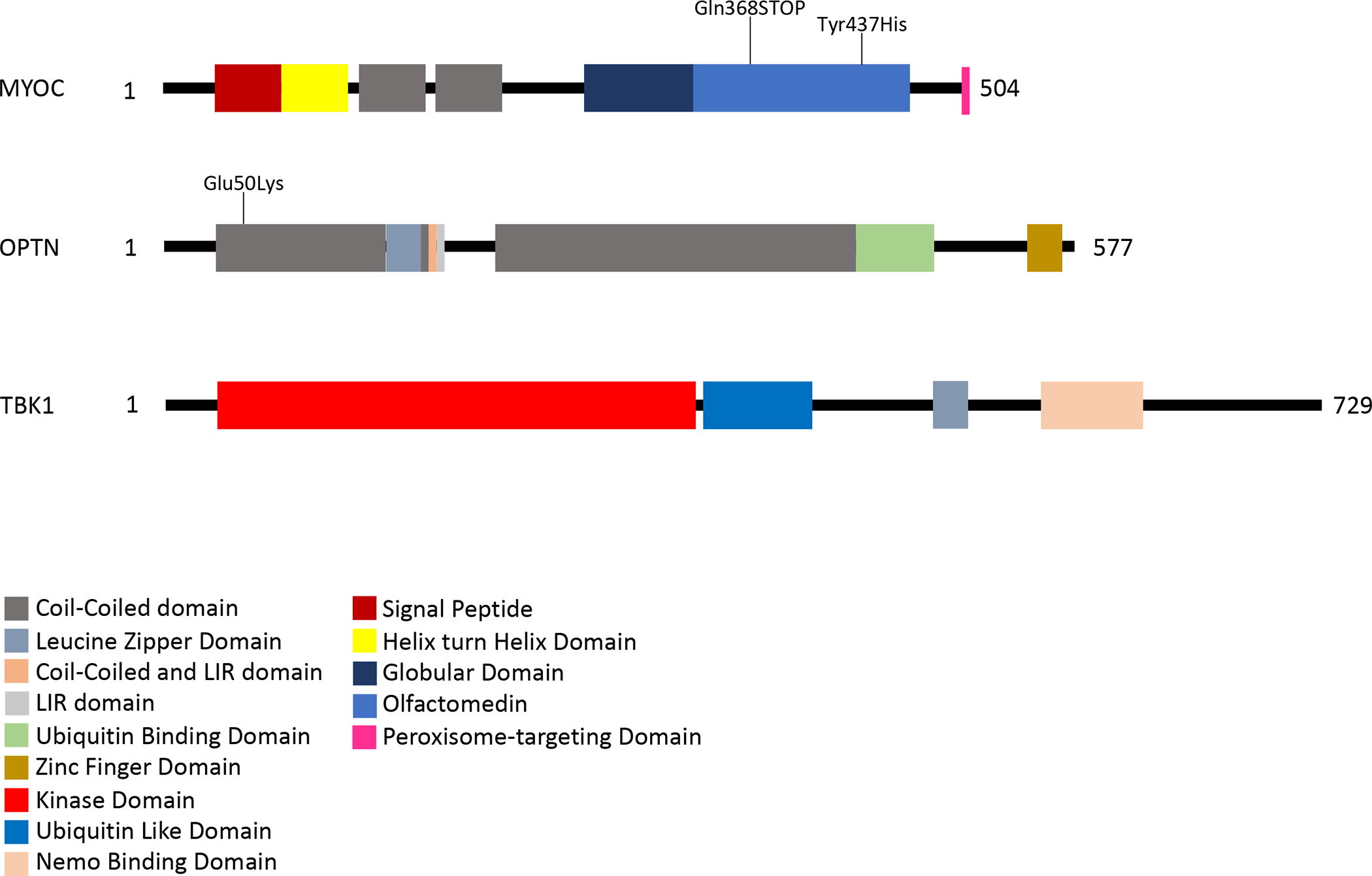
Figure 1. Protein Structure of MYOC, OPTN, and TBK1.

Each protein is represented by a linear diagram proportional to its length in amino acids (AA), 504 AA for MYOC, 577 AA for OPTN, and 729 AA for TBK1. Functional domains and sequence motifs are indicated by colored boxes. The location of some of the more commonly detected glaucoma-causing mutations are indicated on these diagrams.
