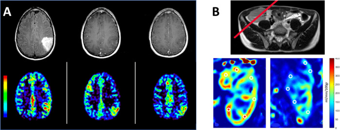
Figure 7.
ASL perfusion MRI from a patient (A) with a brain tumor that is contrast-enhanced in T1 weighted imaging (top row) and increased rCBF (bottom row). Composed from unpublished data. (B) In patients receiving renal transplants, ASL perfusion MRI provides functional and quantitative assessments of the renal filtration, showing a normal blood perfusion map (left) and impaired blood perfusion of the kidney (right). T2 weighted image was used to place the slice (red line) where the labeling RF was applied. Reprinted with permission from ref (80). Copyright 2022 Springer Nature.

