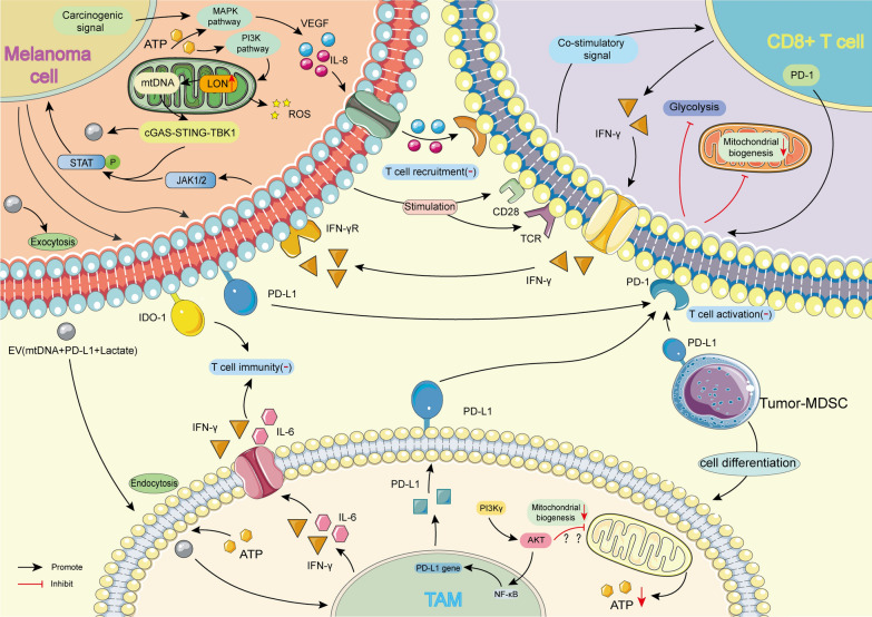
Fig. 4.
Changes of mitochondrial function in different cells during drug resistance of melanoma TME. Melanoma cells, Tumor-MDSCs, and TAMs can all express PD-L1, which makes CD8+ T cells in a resting state. Among them, the expression of PD-L1 in melanoma cells is related to the JAK1/2-STAT pathway involved in mitochondria, while the expression of PD-L1 in TAM may be related to the PI3K γ-NF κ B pathway. When CD8+T cells receive dual-signal stimulation from melanoma cells, they can release IFN- γ to participate in immune regulation. After receiving the extracellular carrier (EV) released by melanoma cells, TAMS also releases interferon-γ and IL-6 to inhibit the immune function of T cells. In addition, melanoma cells can inhibit T cell aggregation by producing VEGF and IL-8 through the MAPK pathway, affecting their mitochondria’s function through the PI3K pathway. Tumor-MDSCs: Tumor-associated myeloid-derived suppressor cells; TAM: Tumor-associated macrophages; IFN- γ: Interferon-gamma; VEGF: Vascular endothelial growth factor; IDO-1: Indoleamine 2,3-Dioxygenase-1; EV: Extracellular vesicle

