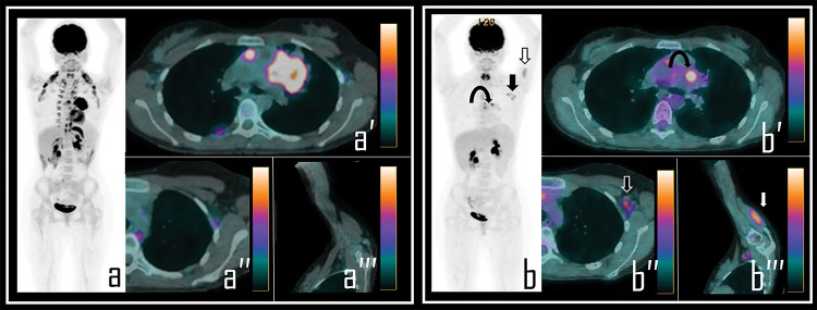
Figure 1. A 36-year-old male patient examined by 2-[18F]FDG PET/CT during the staging of Hodgkin’s lymphoma. Baseline PET/CT (a) showed a mediastinal bulky lymphoma, more evident in axial PET/CT view (a′). No abnormal foci of uptake were observed in left axilla neither in ipsilateral deltoid muscle, as showed in axial PET/CT detail (a′′) and oblique PET/CT view of the left arm (a′′′).
Note: After 2 cycles of chemotherapy, the patient underwent interim 2-[18F]FDG PET/CT, showing incomplete response to therapy in the mediastinal mass with residual lymphoid active tissue, as evident in PET Maximum Intensity Projection (b, curved arrow). PET also showed focal uptake in the left deltoid (b, white arrow) and in three ipsilateral axillary lymph nodes (b, black arrow). Residual lymphoma is evident in axial PET/CT view (b′, curved arrow) while axial PET/CT detail shows a 2-[18F]FDG-avid lymph node in left axilla (b″, black arrow), with SUVmax 2.5; oblique PET/CT view of the left arm displays diffuse uptake in the deltoid muscle (b′′′, white arrow), with SUVmax 2.8. The Patient underwent first dose of COVID-19 vaccine 9 days before the second PET/CT scan.

