. 2023 Apr 10;31(2):117–124. doi: 10.32604/or.2023.027705

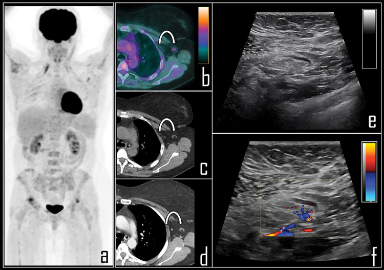
Figure 3. A 65-year-old female patient was examined by means of 2-[18F]FDG PET/CT during follow-up of ovarian cancer. PET Maximum Intensity Projection (a) was negative for disease relapse but showed a focal area of tracer uptake in the left axilla. Patient was previously submitted (14 days before) to second dose of COVID-19 vaccine in the deltoid muscle of left arm. Axial PET/CT view (b, curved arrow) shows the uptake in association with a 0.8 cm wide lymph node, with fatty hilum at correlative low dose axial CT (c, curved arrow). Subsequently, patient underwent whole body contrast enhanced CT, not showing disease relapse but confirming the axillary lymph node in left axilla, with low contrast enhancement (d, curved arrow). Ultrasonography was performed, displaying benign, 0.8 cm wide lymph node with regular shape (e), dominant central hilar flow and peripheral flow at color-doppler (f).

Figure 3