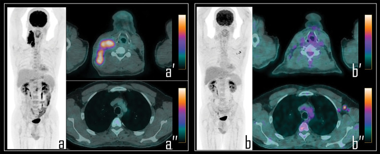
Figure 4. A 25-year-old male patient underwent 2-[18F]FDG PET/CT during the staging of Hodgkin’s lymphoma. PET (a) demonstrated pathologic tracer uptake in right lateral cervical lymphadenopathies. Axial PET/CT view well displays pathologic lymphadenopathies in the neck (a′) while no foci of uptake were detected in the axillary regions (a″). Subsequently, patient was submitted to chemotherapy and, 9 days before the interim PET/CT, to COVID-19 vaccine administration in the left arm.
Note: Interim PET/CT (b) showed response to therapy with reduction of the uptake in the neck, more evident in axial PET/CT view (b″); conversely, three lymph nodes with moderate 2-[18F]FDG uptake were detected in the left axilla, as displayed in axial PET/CT view (b″), presenting SUVmax 2.8. These findings were linked to the previous vaccine administration.

