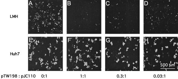
FIG. 2.
Variation in the expression of GFP reveals the toxic effect of delta protein in transfected avian cells. Cultures of LMH cells (A to D) and Huh7 cells (E to H) were cotransfected with fixed amounts of plasmids pGG119 and pJC110 along with variable amounts of pTW198. The ratios of pTW198 to pJC110 are indicated. At 4 days after transfection, fluorescence microscopy, coupled with a charge-coupled device camera, was used to record the levels of GFP fluorescence.

