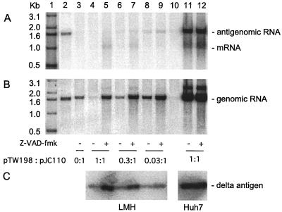
FIG. 5.
Assays of HDV replication in avian and mammalian cells during expression of small delta protein in the presence of the antiapoptotic compound Z-VAD-fmk. Cultures of LMH (lanes 3 to 10) or Huh7 cells (lanes 11 and 12) were cotransfected with fixed amounts of pGG119 and pJC110 along with various amounts of pTW198. The ratios of pTW198 to pJC110 are as follows: lane 3, 0:1; lanes 4, 5, 11, and 12, 1:1; lanes 6 and 7, 0.3:1; lanes 8 and 9, 0.03:1. Lane 1 contains 5′-labeled DNA size markers. Lane 2 is a standard of unit-length HDV cDNA (200 pg). Lane 10 is a sample from untransfected cells. Lanes 5, 7, 9, and 12 correspond to cultures treated, beginning 1 h prior to cotransfection, with 40 μM Z-VAD-fmk. At 4 days after transfection, RNA and protein were extracted. RNA was analyzed by Northern blot for antigenomic RNA (A) or genomic RNA (B). Protein was assayed by immunoblot for delta protein (C).

