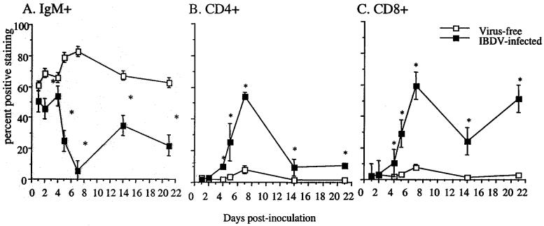
FIG. 1.
Lymphocyte subpopulations in the bursae following IBDV exposure. At 1, 2, 4, 5, 7, 14, and 21 days p.i., bursal cells from virus-free control and IBDV-infected chickens were stained with monoclonal antibodies against chicken μ chain (A), CD4 (B), and CD8 (C). The results presented are the mean of three pools of each group (four chickens per pool) ± SD. Asterisks indicate statistically significant differences between virus-free and virus-exposed groups (P < 0.03).

