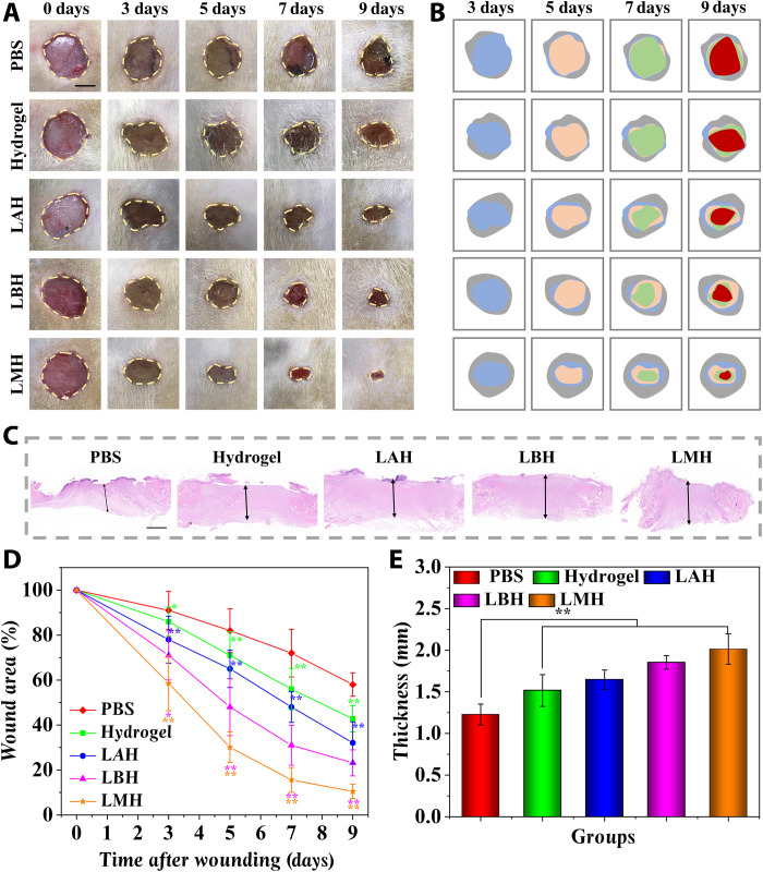
Fig. 5. The LMH accelerated the wound healing process of the infected diabetic wounds.
(A) Representative images of full-thickness skin defect wounds treated by PBS, hydrogel, LAH, LBH, and LMH. (B) Schematic illustrations of the healing process of the wound bed. (C) The regenerated granulation tissues were stained by hematoxylin and eosin (H&E) staining after 9 days. (D) Quantification of the wound area healed by different treatments. (E) Quantification of granulation tissue thickness in different groups. Scale bars, 5 mm (A) and 1 mm (C). n = 6 per group. *P < 0.05 and **P < 0.01.

