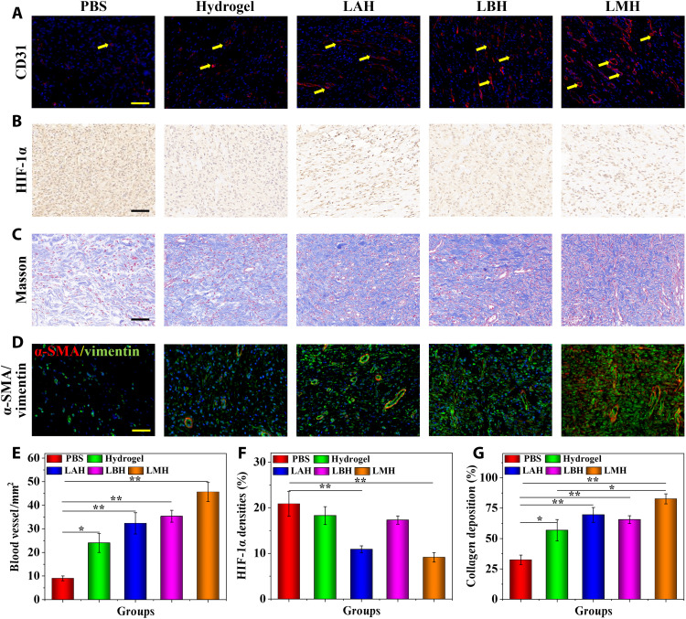
Fig. 6. The mechanism of LMH promoting chronic wound repair.
(A) The CD31-positive blood vessel endothelial cells were stained as red. (B) Immunohistochemical staining of HIF-1α in granulation tissues in different groups. (C) Masson’s trichrome staining of granulation tissues in different treatments. (D) Double immunofluorescence staining of α-SMA and vimentin of granulation tissues in different treatments. (E to G) Quantification of (E) blood vessel number, (F) HIF-1α densities, and (G) collagen deposition. Scale bars, 100 μm (A to D). n = 6 per group. *P < 0.05 and **P < 0.01.

