. 2023 May 11;12:e85041. doi: 10.7554/eLife.85041

Figure 2. Implementation of retro-Tango in the giant fiber and central complex circuits.

(A) Initiating retro-Tango from the GFs (asterisks mark the cell bodies) results in presynaptic signal in the brain and VNC (223±59 neurons in 5 brains, 1±3 neurons in 5 VNCs). Both LC4 (arrow) and LPLC2 (arrowhead) neurons, known presynaptic partners of GFs, are identified by retro-Tango. Note the asymmetry between hemispheres in the signal in the postsynaptic starter neurons and their corresponding presynaptic partners. (B) retro-Tango exhibits little background noise in the absence of a Gal4 driver. Background is observed in the mushroom bodies, in the central complex, and in a few neurons in the VNC (68±10 neurons in 4 brains, 1±1 neurons in 4 VNCs). (C) Ligand expression in EPG neurons of the central complex leads to retro-Tango signal in their known presynaptic partners: PEN, PFR and Δ7 neurons (170±24 neurons in 5 brains). The signal in these neurons can be easily discerned from the background noise. 15do males were analyzed for all panels. Postsynaptic GFP (cyan), presynaptic mtdTomato (magenta) and neuropil (grey). Scale bars, 50 μm.

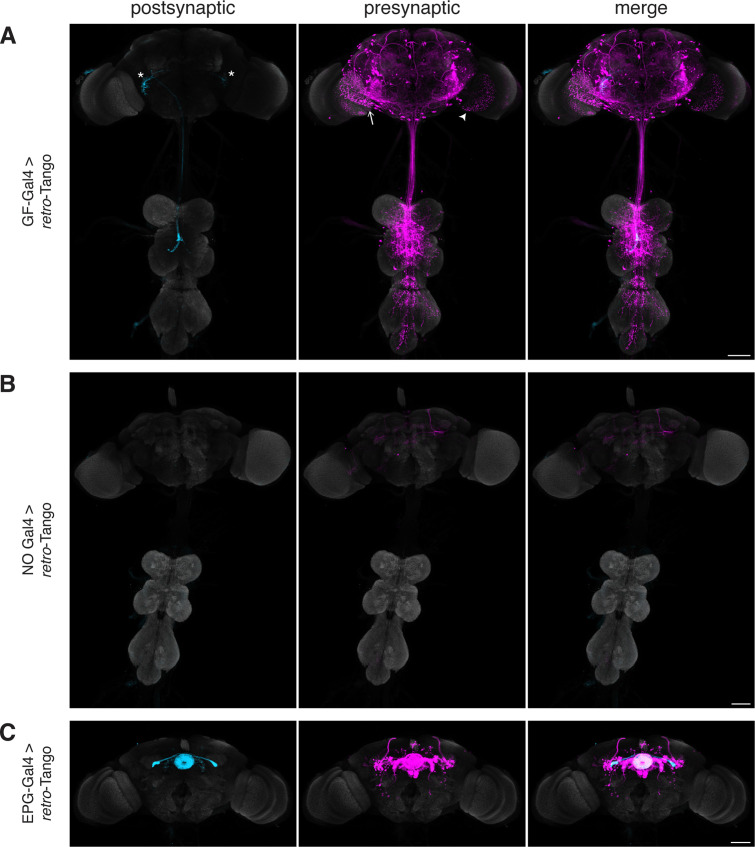
Figure 2.

Figure 2—figure supplement 1. trans-Tango in the giant fiber and central complex circuits.

Figure 2—figure supplement 1.

(A) Initiating trans-Tango from the GFs results in strong postsynaptic signal in the VNC and little to no signal in the brain (4±2 neurons in 4 brains, 48±16 neurons in 4 VNCs). (B) Expression of the trans-Tango ligand in the EPG neurons of the central complex reveals their postsynaptic partners (255±22 neurons in 5 brains). Note the stronger signal in the LAL (arrow) and the weaker signal in the EB (arrowhead) compared to retro-Tango results (Figure 2C). 20do males were analyzed for both panels. Presynaptic GFP (cyan), postsynaptic mtdTomato (magenta) and neuropil (grey). Scale bars, 50 μm. (C) Comparison of the pixel intensities for the signals of retro-Tango and trans-Tango for the EPG circuit in the ellipsoid body (n=5 brains each). (D) Comparison of the pixel intensities for the signals of retro-Tango and trans-Tango for the EPG circuit in the lateral accessory lobes (n=10 hemibrains each). Dots represent data points, the horizonal lines represent the mean and the error bars represent the standard error of the mean. Student’s t-test, *: p<0.05, ****: p<0.0001.
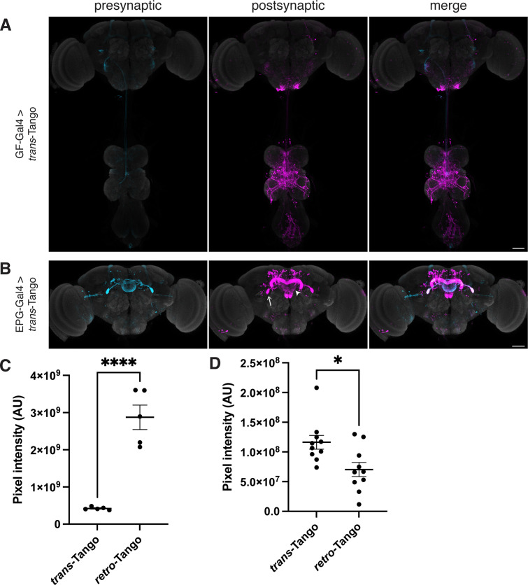
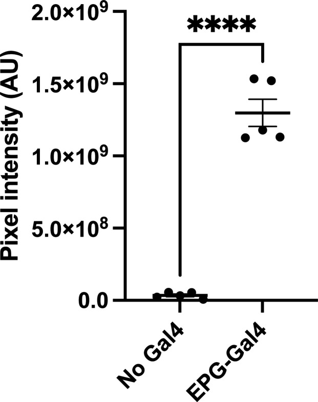
Figure 2—figure supplement 2. The retro-Tango signal in the EPG circuit is far stronger than the background noise.

Figure 2—figure supplement 2.

Comparison of the pixel intensities in the central complex for the background noise signal of retro-Tango and retro-Tango signal when initiated from EPG neurons (n=5 brains each). Dots represent data points, the horizonal lines represent the mean and the error bars represent the standard error of the mean. Student’s t-test, ****: p<0.0001.
Figure 2—figure supplement 3. retro-Tango does not yield false positive signal in neighboring neurons in the EPG circuit.

Figure 2—figure supplement 3.

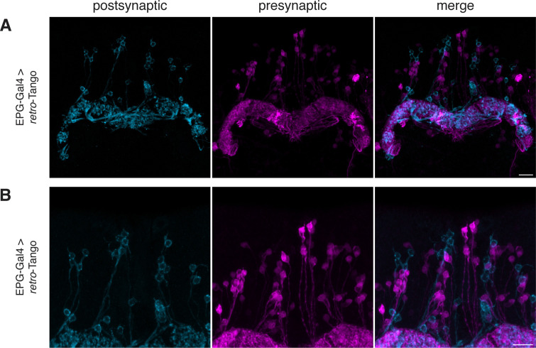
(A–B) When retro-Tango is initiated from EPG neurons, the ligand present in the cell bodies does not lead to false positive presynaptic signal in neighboring neurons. For clarity, only a subset of the z-stack projection is shown. 15do males were analyzed. Postsynaptic GFP (cyan), presynaptic mtdTomato (magenta). Scale bars, 10 μm.
Figure 2—video 1. retro-Tango does not yield false positive signal in neighboring neurons in the EPG circuit.
Download video file (786.5KB, mp4)
Video through the z-stack sections of the image in Figure 2—figure supplement 3B. Postsynaptic GFP (cyan), presynaptic mtdTomato (magenta).