Fig. 2.
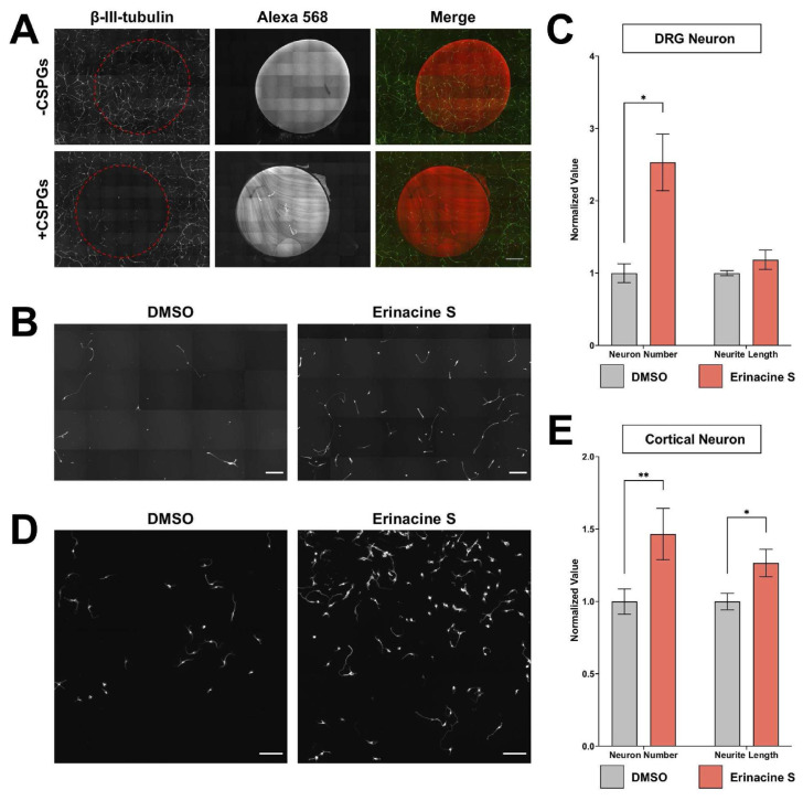
Erinacine S promotes cortical neuron regeneration on inhibitory substrates. (A) Dissociated rat DRG neurons cultured on surface with or without CSPGs. Alexa Fluor 568-conjugated antibody was mixed with ddH2O (top) or CSPGs (bottom) to label the coated region. Neurons were immunofluorescence stained with the antibody against the neuron-specific β-III tubulin (green). The red dotted area indicates ddH2O or CSPGs-coated region. All images have the same scale and the scale bar represents 1 mm. (B) Representative images of dissociated rat DRG neurons cultured in the CSPGs-coated area and treated with 1 μg/mL erinacine S or DMSO solvent control. Neurons were immunofluorescence stained with the antibody against neuron-specific β-III tubulin. Both scale bars represent 300 μm. (C) Quantification of DRG neuron number (left) and total neurite length per neuron (right) from 4 independent experiments in CSPGs-coated regions. *p < 0.05, two-tailed Student's t-test. (D) Representative images of dissociated mouse cortical neurons cultured in the CSPGs-coated area and treated with 1 μg/mL erinacine S or DMSO solvent control. Neurons were immunofluorescence stained with the antibody against neuron-specific β-III tubulin. Both scale bars represent 100 μm. (E) Quantification of cortical neuron number (left) and total neurite length per neuron (right) from 4 independent experiments in CSPGs-coated regions. *p < 0.05, **p < 0.005, two-tailed Student's t-test.

