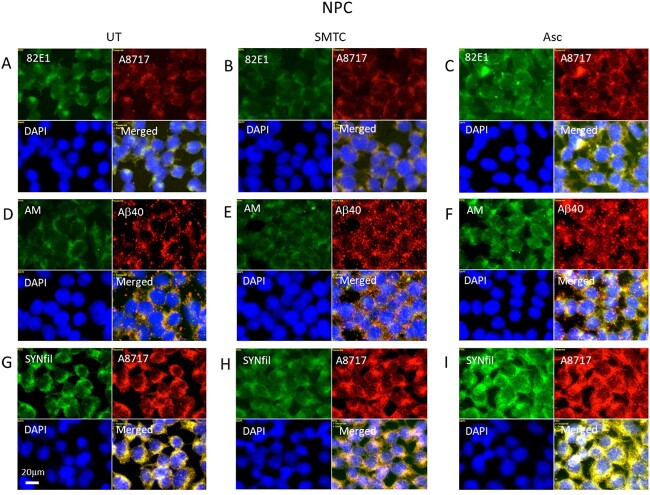
Fig. 4.
Modulation of APP and GPC1 processing and SYNfil formation. Representative immunofluorescence images of cells that were grown to confluence in A, D, G) regular medium (UT, untreated) or in B, E, H) medium containing100 μM SMTC or in C, F, I) medium containing 1 mM ascorbate (Asc). Staining was performed with mAb 82E1 (for the N-terminal of β-CTF/Αβ, green), mAb AM (for HS-anMan, green), mAb SYNfil (green), pAb A8717 (for the C-terminal of APP/β-CTF, red), pAb Aβ40 (for the Aβ region of APP/β-CTF, red), and DAPI (for nuclei, blue). Exposure time was the same in all cases. Bar, 20 μm.

