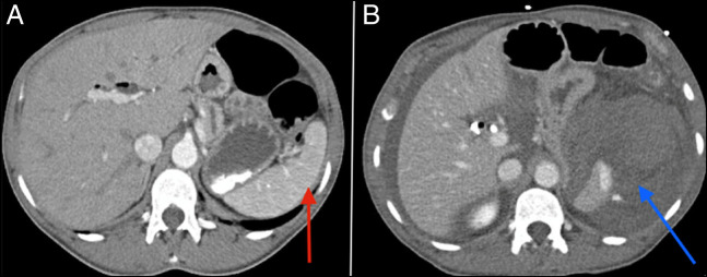
Figure 4.
(A) A late arterial phase abdominal CT scan showing a normal spleen (red arrow). (B) A portal venous phase abdominal CT scan showing large perisplenic hematoma and hemoperitoneum with arterial extravasation of contrast consistent with active arterial bleeding (blue arrow). CT, computed tomography.

