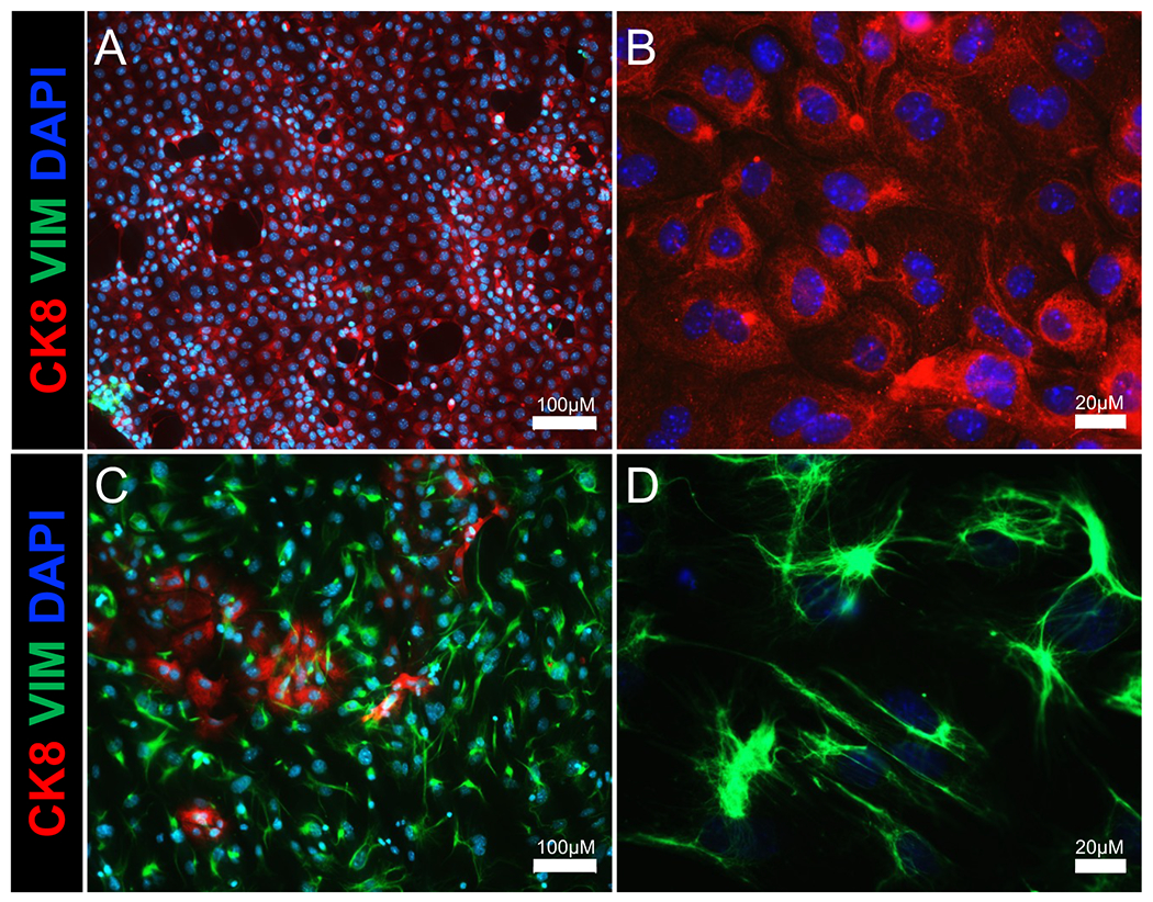
Figure 2: Immunostaining of isolated endometrial epithelium and stromal cell populations.

Immunofluorescence images show epithelial and stromal cell populations of the (A,B) epithelial cell and (C,D) stromal cell fractions following enzymatic and mechanical separation of the uterus. Cytokeratin 8 (an epithelial cell marker) is shown in red, vimentin (a stromal cell marker) is shown in green, and DAPI (a nuclear marker) is shown in blue. Scale bars = 100 μm (A,C), 20 μm (B,D). Abbreviations: CK8 = cytokeratin 8; VIM = vimentin; DAPI = 4′,6-diamidino-2-phenylindole.
