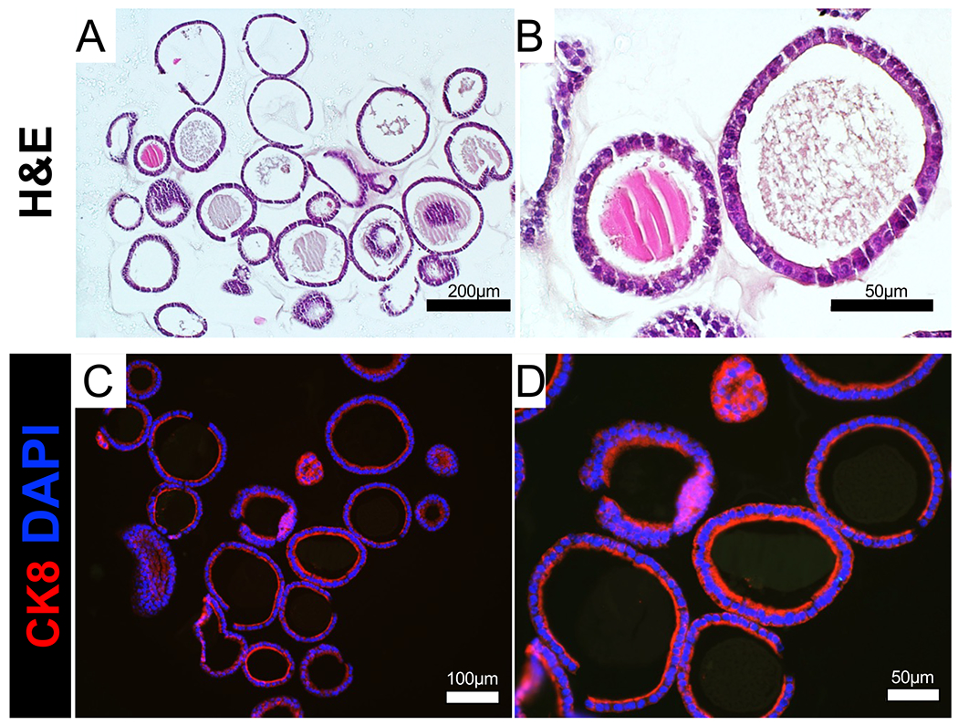
Figure 4: Histological analysis of endometrial organoids.

(A,B) FFPE endometrial epithelial organoids were sectioned and stained with H&E. (C,D) FFPE epithelial organoids were immunostained with the epithelial cell marker cytokeratin 8 (red). Cell nuclei were stained with DAPI (blue). Scale bars = 200 μm (A), 100 μm (C), 50 μm (B,D). Abbreviations: FFPE = Formalin-fixed paraffin embedded; H&E = hematoxylin & eosin; CK8 = cytokeratin 8; DAPI = 4′,6-diamidino-2-phenylindole.
