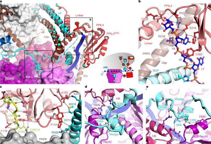
Fig. 4. PRP2 translocation terminates in a 3D construction reminiscent of a molecular brake.
a, Interactions between the subunits of the molecular brake. Interacting residues are shown as spheres depicted in the same colour as their binding partner. The domains of PRP2 RecA1, RecA2, HB and NTD are depicted. The anchoring surfaces of the brake elements to PRP8 (grey oval) are indicated. The inset shows a schematic representation of the molecular brake, in which the subunits are coloured as in the surface and cartoon representation. CWC15 is yellow. b, Interactions of the intron with PRP2NTD, PPIL4 and SKIP. c, Interactions of PPIL4 with CWC15 and SKIP. d,e, The wedge element of SKIP intercalates between RecA1, RecA2 and the HB domains of PRP2, indicating inhibition of translocation. PPIL4, SKIP and CWC15 are red, cyan and yellow, respectively.

