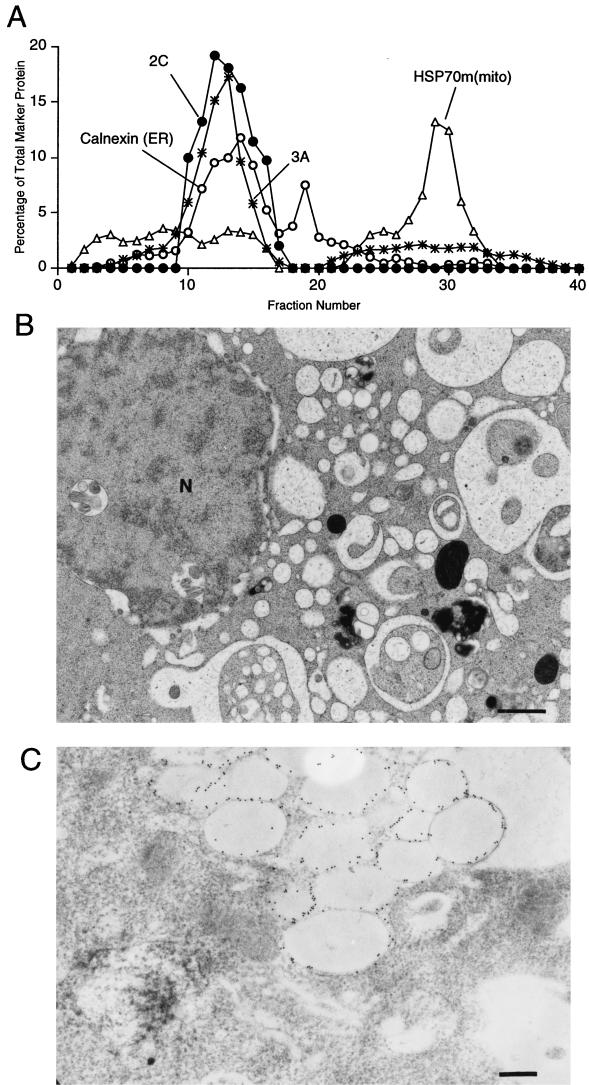
FIG. 4.
Density gradient and ultrastructural analysis of COS-1 cells that expressed poliovirus proteins 2C and 3A. (A) Cytoplasmic membranes from cells expressing 2C and 3A proteins were separated on a 12% stock isotonic Percoll gradient; proteins in each fraction were detected by immunoblotting using antibodies directed against poliovirus 2C, poliovirus 3A, calnexin (ER), and mtHSP70 [HSP70m(mito) mitochondria]; lightest fractions are at the left. (B) Electron microscopy reveals the effects of 2C and 3A expression on the ultrastructure of COS-1 cells. N, nucleus. Bar = 1 μm. (C) Section labeled with 15-nm gold particles coupled to secondary antibodies. The primary antibody was directed against poliovirus protein 2C. Bar = 0.5 μm.

