Figure 6.
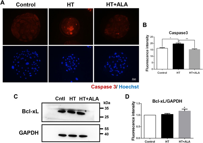
Effect of ALA on apoptosis in heat stressed-porcine parthenotes. (A) Representative images of caspase3 after HT exposure in presence of ALA in porcine parthenotes. Red, caspase3; Blue, DNA. (B) Relative fluorescence intensity of caspase3 after HT exposure with ALA addition. (C) Western blotting of Bcl-xL protein expression after HT exposure with ALA treatment. Original blots are presented in Supplementary Fig. 1B′,C. (D) Relative band intensity analysis for Bcl-xL/GAPDH after HT exposure with ALA supplementation. *p < 0.05; **p < 0.01.

