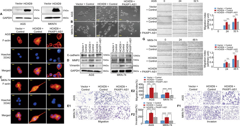
Fig. 4. HOXD9-PAXIP1-AS1 axis regulates the migration and invasion of GC cells in vitro.
A The efficiency of HOXD9 plasmid transfection was detected by western blotting assay. B The morphology of GC cells was observed under phase-contrast microscopy. C Transfected AGS and MKN-74 cells were stained with rhodamine-phallotoxin to identify F-actin filaments under the fluorescent microscope. All experiments were repeated three times. D EMT biomarkers, including E-cadherin, MMP2, and Vimentin were detected by western blotting 48 h after transfection. E, F Transwell migration (E1) and invasion (F1) assay were performed on GC cells. Quantification of migration and invasion capabilities of GC cells are shown (E2, F2). ***P < 0.01, ****P < 0.001. G The wound healing assay was used to detect cell motility of AGS and MKN-74 cells following transfection. The migration index is shown in the right panel. **P < 0.01, ***P < 0.01, ****P < 0.001. Scale bars, 25 μm in (B) and 10 μm in (C).

