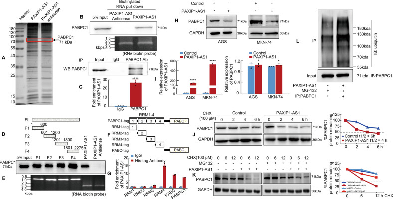
Fig. 5. PAXIP1-AS1 binds to Polyadenylate-binding protein cytoplasmic 1 (PABPC1) in human GC cells.
A Silver staining of biotinylated PAXIP1-AS1-associated proteins. B Western blotting of proteins from PAXIP1-AS1 and its antisense pull-down assays; n = 3. C The association of endogenous PABPC1 and PAXIP1-AS1 in MKN-74 cells was detected by RNA immunoprecipitation (RIP) assay using anti-PABPC1 antibodies. Relative enrichment (mean ± SD) of PAXIP1-AS1 with anti-PABPC1 or anti-IgG was measured by qPCR. D, E Western blotting of PABPC1 in samples pulled down by full-length or truncated PAXIP1-AS1 (F1: 1–600, F3: 601–1200, F3: 1201–1800, F4: 1801–2275); n = 3. F, G Full-length and truncated PABPC1 plasmids with 6× His-tag were transfected into MKN-74 cells for 48 h and extracted for RIP assays with anti-His antibody. Results showed that PAXIP1-AS1 mainly bound to the RRM4 region and PABC domain in full-length PABPC1. H Western blotting analysis of PABPC1 in Control and PAXIP1-AS1-overexpressed GC cells. I Total RNA from Control or PAXIP1-AS1-overexpressing AGS and MKN-74 cells was analysed by qPCR to validate PAXIP1-AS1 expression (the left panel) and determine PABPC1 mRNA levels (the right panel). ****P < 0.001 and *P > 0.05, Control vs. PAXIP1-AS1. n = 3. J Control or PAXIP1-AS1-overexpressed MKN-74 cells were treated with 100 μM CHX for indicated periods. Subsequently, cell lysates were analysed by western blotting with anti-PABPC1 and anti-GAPDH antibodies. K PAXIP1-AS1 or control plasmid was transfected into MKN-74 cells for 48 h and cells were treated with DMSO, CHX (100 μM), or CHX plus MG132 (10 nM) for 0, 6, or 12 h before protein extraction. Subsequently, cell lysates were analysed by western blotting with anti-PABPC1 and anti-GAPDH antibodies. L PAXIP1-AS1-overexpressed or control plasmid was transfected into MKN-74 cells for 48 h and cells were treated with 10 nM MG132 for 6 h. Extracts were immunoprecipitated with anti-PABPC1 antibody, and PABPC1 polyubiquitination was examined by western blotting using anti-ubiquitin antibody.

