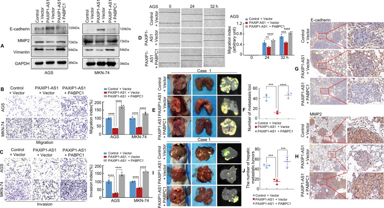
Fig. 6. PAXIP1-AS1 with PABPC1 regulates migration and invasion of GC cells.
A The expression of EMT biomarkers, including E-cadherin, MMP2, and Vimentin was detected by western blotting 48 h following transfection. B, C GC cells were transfected and subjected to Transwell migration and invasion assay. Quantification of migration and invasion capabilities of GC cells are shown in the right panel. ****P < 0.001. D The mobility of transfected AGS cells was detected by the wound healing assay and the migration index is shown in the right panel. **P < 0.05, ***P < 0.01, ****P < 0.001. E Representative images of metastatic tumours in the lungs from mice in the Control + Vector, PAXIP1-AS1 + Vector, and PAXIP1-AS1 + PABPC1 groups are shown, respectively (n = 3 in each group). F The number of metastatic tumours in the lung was counted. ***P < 0.001, ****P < 0.001. G, H IHC staining of E-cadherin and MMP2 expressions. I, J Representative images (I) and qualification (J) of hepatic metastatic tumours in the indicated groups (n = 3). ***P < 0.01. Scale bar, 100 μm in (G) and (H).

