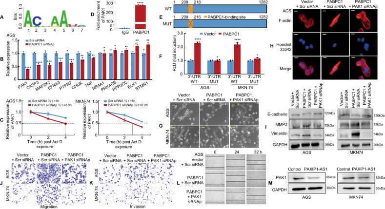
Fig. 7. PABPC1 promotes GC metastasis by enhancing PAK1 mRNA stability.
A PABPC1-binding motif predicted by bioinformatics analysis. B The relative expression of 12 genes in PABPC1 knockdown AGS cells was detected by qPCR. The experiment was performed in triplicate. *P > 0.05; **P < 0.05; ***P < 0.01; ****P < 0.001. Scr siRNA vs. PABPC1 siRNAp. C Actinomycin D (10 μg/mL) was applied to GC cells transfected with PABPC1 siRNAp or Scr siRNA. RNA was extracted at indicated time points to detect the mRNA level of PAK1. D RIP assay in MKN-74 cells showed that PABPC1 bound directly to PAK1 mRNA; anti-IgG was used as a negative control. ****P < 0.001. IgG vs. PABPC1. E The PAK1 3′UTR region is shown in the blue box. The orange box represents a PABPC1 potential binding site(WT, upper panel) and the orange box with a cross represents the corresponding mutation site(MUT, lower panel). F PABPC1 overexpression enhanced luciferase activity in GC cells transfected with the PAK1 3′UTR wild-type reporter construct as compared to those transfected with the mutant reporter construct. *P > 0.05; ****P < 0.001. Vector vs. PABPC1. G The morphology of transfected AGS and MKN-74 cells was observed under phase-contrast microscopy. H Transfected AGS cells were stained with rhodamine-phallotoxin to visualise F-actin filaments under the fluorescence microscope. I PAK1 knockdown decreased the expression of mesenchymal markers and restored the expression of epithelial markers in PABPC1 overexpressed GC cells. J, K GC cells were transfected and subjected to Transwell migration and invasion assays. L The wound healing assay was used to detect GC cell motility. M Control and PAXIP1-AS1 were transfected into GC cells. PAK1 expression and GAPDH were detected in GC cells using western blotting. Scale bars, 25 μm in (G) and 10 μm in (H). For quantification, see Supplementary Fig. 8.

