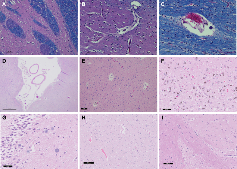
Figure 3.
Routinely histology stainings of tissue sections from perfused brains
Representative histology sections were luxol fast blue, hematoxylin and eosin (LH&E)-stained in case 1 (A-C) or hematoxylin and eosin (H&E)-stained from case 2 (D), case 3 (E), case 4 (F-G), and case 5 (H-I). Sections were derived from the striatum (A-B), corpus callosum (C), temporal cortex (D), white matter of the temporal pole (E), substantia nigra (F), hippocampus – dentate gyrus (G), mid-frontal cortex (H), and striatum (I). Tissue morphology shows expected findings, suggesting that the perfusion process did not adversely affect tissue morphology. Red blood cells were observed in some vessels, indicating absence of complete perfusion. All scale bars 100 μm except for sub-figures D (1000 μm), G (50 μm), and H (200 μm).

