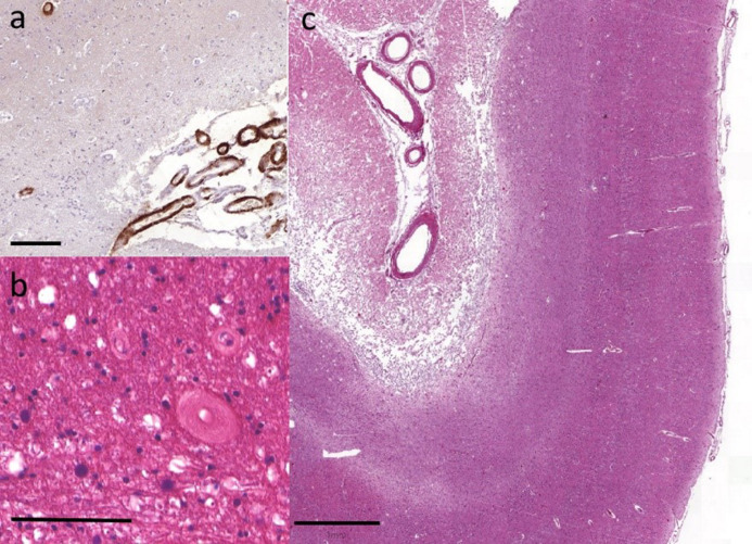
Figure 1. Representative cerebrovascular lesions.
Moderate to severe (a) leptomeningeal CAA was frequently observed in the occipital cortex. (b) Severe white matter arteriolosclerosis was not uncommon particularly in the occipital white matter adjacent to the ventricle. (c) Infarcts were observed in all subcortical and cortical regions, including this large infarct affecting occipital cortex. Scale bar is 100um in (a) and (b) and 1mm in (c).

