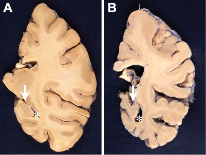
Figure 1.

Comparison between formalin-fixed brain slices of the left hemispheres (level of posterior hippocampus) of an aged nondemented individual (A) and an AD patient (B). Note the marked atrophy (thinning of the gyri and deepening of the sulci) in B, in particular hippocampal atrophy (arrow in B) with widening of the inferior horn of the second ventricle (asterisk in B). Photographs by courtesy of Simon Fraser and Arthur Oakley.
