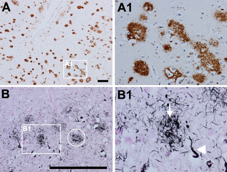
Figure 2.
Amyloid and neuritic plaques. A; A1. Multiple diffuse amyloid plaques in the neocortex (antibody 4G8). B, B1. Neuritic plaques that contain Aβ and tau in distended processes (i.e. dystrophic neurites). Gallyas silver stain visualizes both aggregated Aβ and tau and is therefore ideal to detect neuritic plaques (ring in B, neuritic plaque; arrow in B1, dystrophic neurite; arrowhead in B1, neurofibrillary tangle). Scale bars: 200 μm. From [71].

