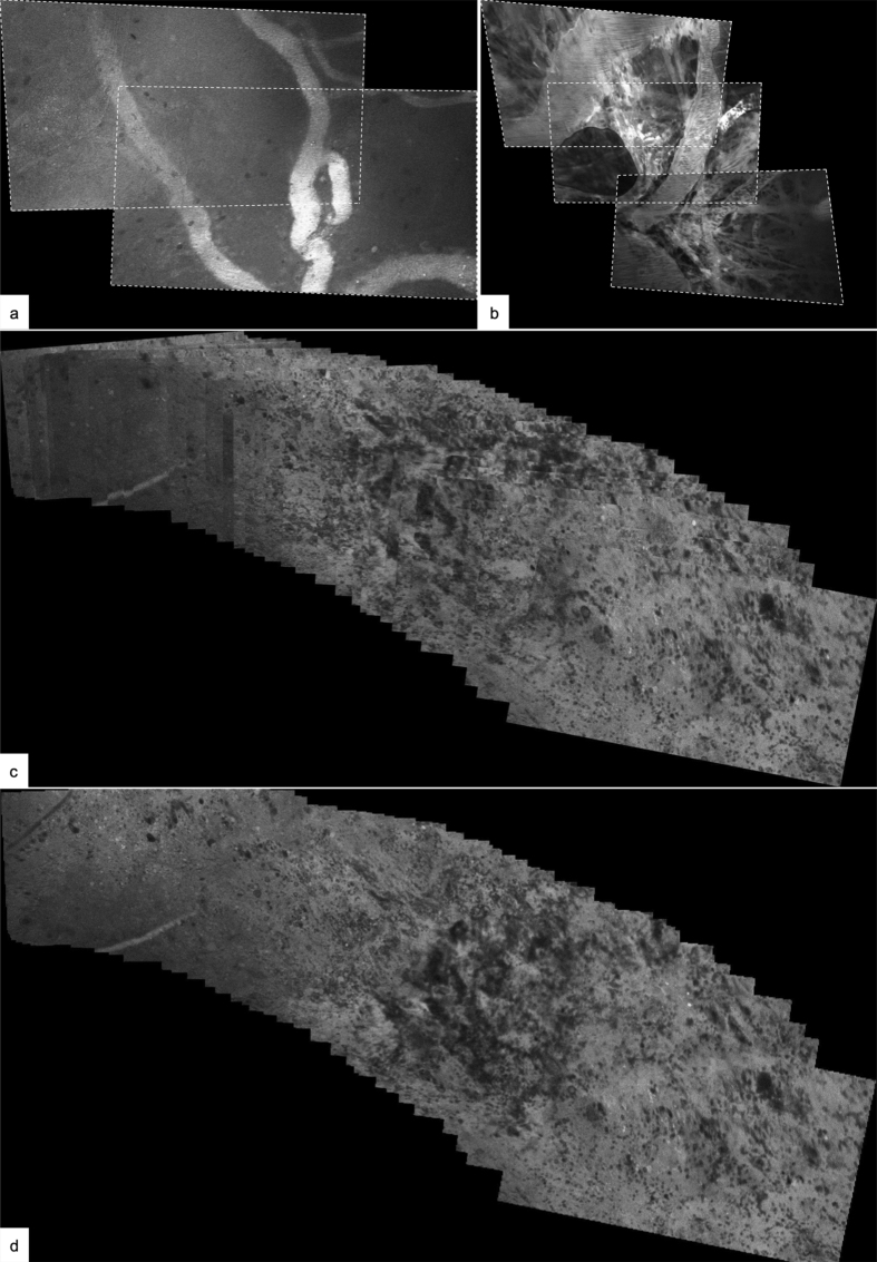
Figure 5.
Experimental image processing (a,b) using stitching techniques. Serial images with overlapping content can be stitched together and result in a greater field of view. Illustrative example of two (a) and three (b) serial images of vasculature with overlapping image content. The images were extracted from an image series recorded over several seconds during which the CLE probe was moved along the tissue surface. Tilting of the probe tip or tissue compression during image series acquisition can result in perspective distortion, which has been corrected for in these examples. Composite of 45 serial images of tumor tissue (c, d): The CLE probe was maneuvered along the tissue surface while serial images were acquired. The images were aligned based on overlapping content (c), which results in a much greater field of view compared to the individual images. Because the images are stacked onto each other, the borders of the individual images are clearly recognizable, as the images on top of the stack overlay the images further below. Further image processing methods, such as blending, can create a seamless tissue map and allow assessment of the tissue in its continuity (d). For clinical purposes it has to be made sure that such methods do not introduce diagnostically relevant artifacts.

