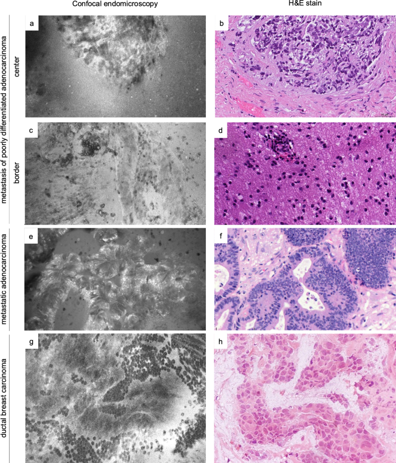
Figure 6.
Comparative examples of metastases in in vivo and ex vivo CLE images and H&E slides. Shown are a metastasis of a poorly differentiated carcinoma (a-b) and reactive changes (gliosis and discrete lymphocytic inflammatory infiltrate) at the border of the same lesion (c-d), metastatic adenocarcinoma (e-f) and metastasis of ductal breast carcinoma (g-h, H&E of frozen section). For illustrative purposes, both in vivo and ex vivo CLE images are included showing that morphology is represented consistently independent of the acquisition modality (a, c, and e: ex vivo; g: in vivo).

