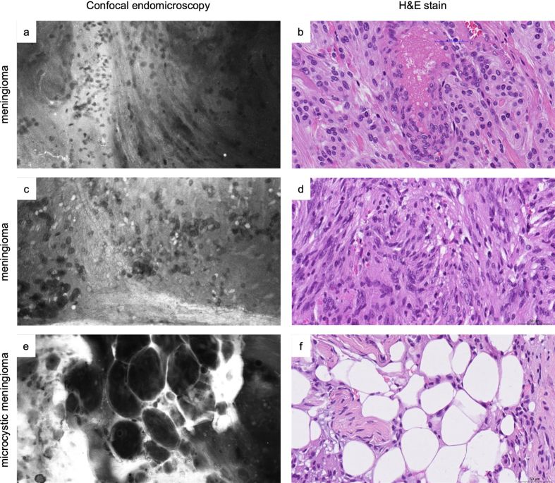
Figure 7.
Comparative examples of tumor morphology in ex vivo CLE images and matched H&E slides in two distinct cases of meningioma (a-b and c-d) and microcystic meningioma (e-f). While in vivo imaging is the preferred usage scenario of CLE, ex vivo imaging is useful for training purposes. It enables recording images over a longer period than what is practicable during surgery. It can also help to point out specific tissue characteristics that might otherwise be more time-consuming to achieve intraoperatively.

