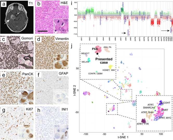
Figure 1. Radiology, histopathology and epigenetic analysis.
a) Representative axial MRI demonstrating the preoperative finding of a large temporomesial tumor (large arrow). Contrast enhancement was found partially within the tumor and additionally in the region of the infundibulum (small arrow). T1-weighted image plus contrast medium.
b) Histomorphology revealed geographical necrosis, prominent rhabdoid morphology and brisk mitotic activity of tumor cells (inset).
c) The tumor showed a prominent network of reticular fibres, visible in the Gomori silver impregnation.
d) Immunostaining for vimentin was vastly strongly positive.
e) Immunostaining for cytokeratins (AE1/3) was vastly strongly positive in the majority of tumor cells.
f) Immunostaining for GFAP was negative in tumor cells while demarcating residual brain tissue.
g) Ki67 proliferative index amounted to about 40%.
h) Tumor cells exhibited loss of nuclear SMARCB1/INI1 staining. Retained nuclear staining was found in blood vessels and inflammatory cells (inset). Scale bar in b – h is 150 µm.
i) Copy number profile of the tumor indicated a homozygous CDKN2A/B (short arrow) and SMARCB1/INI1 (long arrow) deletion.
j) tSNE including a reference set of brain tumors (GSE90496, [11]) showed affiliation of the tumor to the group of PXA.
Clicking the figure will lead you to the full virtual slide (H&E).

