Figure 1.
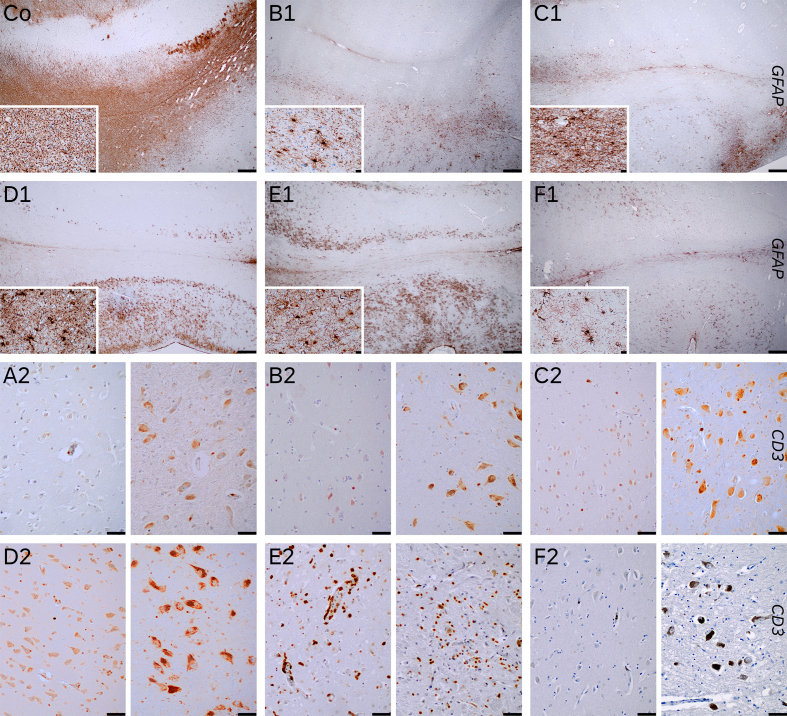
Figures Co to F1: GFAP immunohistochemistry of hippocampus (upper third CA1 sector -right side- and subiculum – left side-) and parahippocampal area (lower third, transenthorinal cortex) of a control (Co) and cases 2 (B1), 3 (C1), 4 (D1), 5 (E1) and 6 (F1). The control was a 52-year-old male who died due to an acute bilateral adrenal hemorrhage with extensive clasmatodendrosis (insets, parahippocampal white matter, 400x). The cases showed variable gliosis, predominantly mild, higher in case 5 (E1) who had an encephalitis, and case 4 (D1) who died with an extensive acute pneumonia. Both had mild and isolated clasmatodendritic changes in white matter (see insets).
Row 3 and 4 show T-cell infiltrates (CD3+) in parahippocampal cortex (left half) and substantia nigra pars compacta (right half), in cases 1 (A2), 2 (B2), 3 (C2), 4 (D2), 5 (E2) and 6 (F2). Only case 5 (E2) presented marked infiltrates, while other cases has only scattered to isolated perivascular T-cells. Case 2 (B2), which was positive for CART PCR in brain tissue, did not have significantly higher T-cell infiltrates (Figures Co, B1 to F1 20x scale bar 500 µm; insets 400x, scale bar 20 µm. Figures A2 to F2 200x, scale bar 50 µm).

