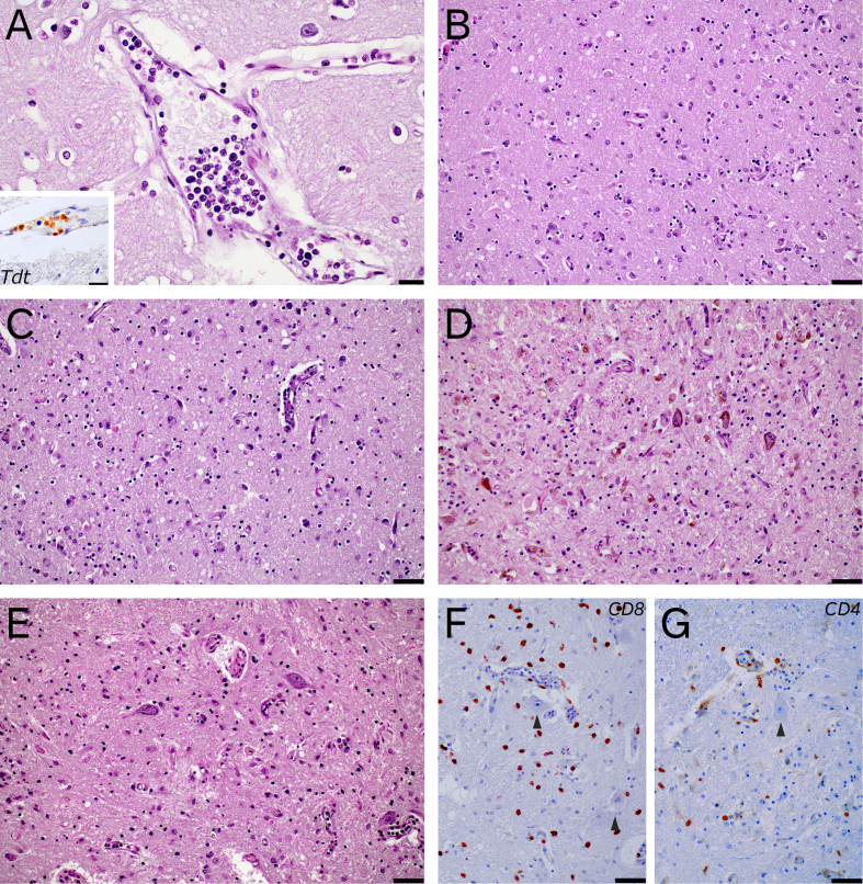
Figure 2.
Remarkable histological features: case 1 presented scant intravascular infiltration of the leukemia (A), which was positive for TdT (A, inset). Case 5 showed an encephalomyelitis, with predominant involvement of limbic areas (B, entorhinal cortex), basal ganglia (C, hypothalamus), brainstem (D, substantia nigra) and spinal cord (E, lumbar spinal cord). T-cell lymphocytic infiltrates were both CD8+ and CD4+, with predominance of the former (F and G, CD8+ and CD4+ infiltrates in lumbar spinal cord, which did not show marked tropism for motor neurons (arrowheads)) (A 400x, scale bar 20 µm, inset 600x, scale bar 20 µm; B to G, 200x, scale bar 50 µm).

