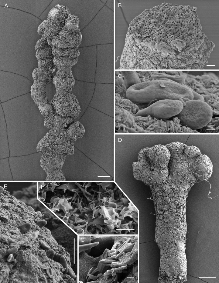
Figure 9.
Scanning electron micrographs of Chaenothecopsisnodosa sp. nov. (PDD 110745) A branched ascoma with numerous tightly stacked capitula B cross section of stipe C ascospore ornamentation D compound capitula E–G details of capitulum surface E ascospores on capitulum surface F amorphous material on capitulum surface G crystals on capitulum surface. Scale bars: 100 µm (A, D); 10 µm (B, E); 1 µm (C, F, G).

