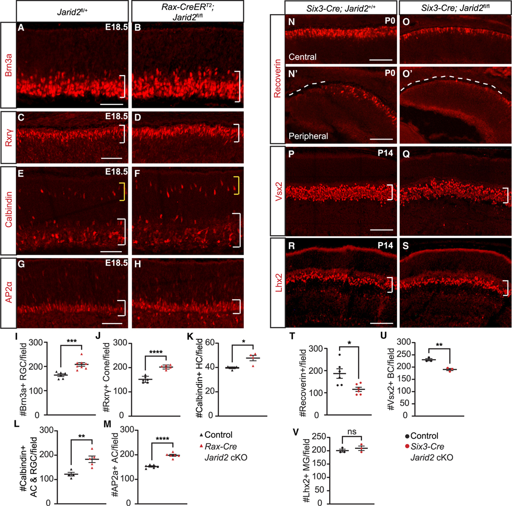
Figure 1. Loss of Jarid2 affects retinal neurogenesis.
Rax-Cre Jarid2 cKO retina at E18.5 shows (A, B, and I) increased Brn3a-labeled RGCs within the GCL (bracket), (C, D, and J) increased Rxrγ-labeled cone photoreceptors (bracket), (E and F) increased calbindin-labeled horizontal cells (yellow bracket, K) and amacrine cells (white bracket, L), and (G, H, and M) increased AP2α-labeled amacrine cells (bracket). At P0, Six3-Cre Jarid2 cKO shows (N, O, and T) reduced Recoverin-labeled photoreceptors in the central retina (N′ and O′) and delayed differentiation in the peripheral retina (dotted line). At P14 (P, Q, and U), Vsx2-labeled bipolar cells were reduced, (R, S and V) but Lhx2-labeled Müller glia were unchanged. Graphs represent mean ± SEM. n = 6 retinas in (J, M, and T) and control in (I), n = 8 for Jarid2 cKO in (I), n = 4 in (K), and control in (L), n = 5 for Jarid2 cKO in (L), and n = 3 in (U and V). *p < 0.05, **p < 0.01, ***p < 0.001, ****p < 0.0001 by Student’s t test. Scale bars, 100 μm. See also Figure S2 and Table S6 for statistical details.

