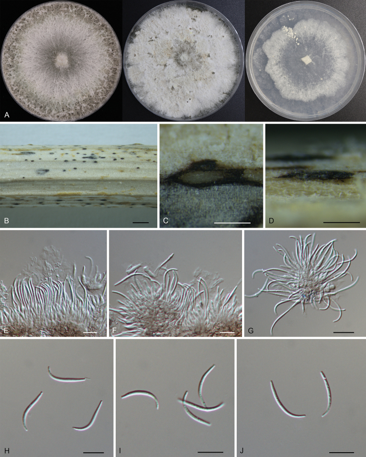
Figure 3.
Morphology of DiaportherizhaoensisA colonies on PDA, MEA and SNA at 25 °C after 2 weeks B habit of conidiomata on the host C transverse section of the conidioma D longitudinal section through the conidioma E–G conidiogenous cells with attached beta conidia H–J beta conidia. Scale bars: 500 µm (B); 100 µm (C, D); 10 µm (E–J).

