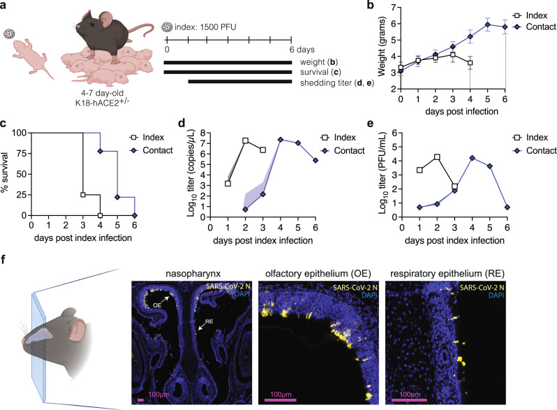
Fig. 1. Establishment of a neonatal K18-hACE2 mouse transmission model using SARS-CoV-2 WA-1.
a Four to seven-day-old K18-hACE2+/- pups were intranasally infected with 1500 PFU of SARS-CoV-2 WA-1 and cohoused with uninfected littermates for 6 days. Weight and survival were monitored daily, and viral shedding samples were collected by dipping the nares of each individual pup in viral medium daily. Data from two independent repetitions, with a total of n = 4 index and n = 9 contact mice. Mean body weight (b) and survival (c) of index and contact pups. Viral burden in shedding samples analyzed by RT-qPCR for SARS-CoV-2 RNA (d) and by plaque assay for infectious virus (e). Data shown as geometric mean (line) with geometric standard deviation (shaded area). Individual values below the limit of detection (50 PFU/ml) were set to 5. f Immunohistochemistry for SARS-CoV-2 N protein in 4-7 day old mice nasopharynx. Pups were infected intranasally with 1500 PFU of SARS-CoV-2 WA-1, and heads were fixed at 2 dpi, paraffin embedded, sectioned through the nasopharynx, and stained for SARS-CoV-2 N protein (yellow) and DAPI (blue) for nuclei. Arrows represent areas magnified in the adjacent panels. OE and RE indicate olfactory epithelium and respiratory epithelium, respectively. Created with BioRender.com.

