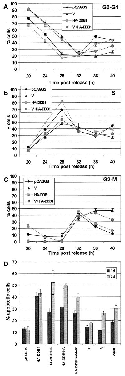
FIG. 7.
Cell cycle analysis on coexpression of V and
HA-DDB1 proteins. Cells were synchronized as described in the legend
for Fig. 2. After the release from the mimosine block, the cells were
transfected with pCAGGS, 2 μg of pCAGGS-V, 3 μg of pCAGGS-HA-DDB1,
or 2 μg of pCAGGS-V plus 3 μg of pCAGGS-HA-DDB1. Additional pCAGGS
DNA was added to the transfections if necessary to ensure that 5 μg
of DNA was present in each transfection. The cells were harvested,
fixed, and permeabilized. The cells were stained with MAb P-k for the
V-expressing cells or with MAb 12CA5, which recognizes the HA-epitope
tag of the cells expressing HA-DDB1. The cells were stained with
propidium iodide and analyzed by flow cytometry as described above. The
high-expressing population of the V-expressing cells was selected for
analysis. The percentages of cells in the G0-G1
(A), S phase (B), and G2-M phase (C) are shown. Each time
point is the average of three plates of cells. Symbols: ⧫,
vector-only-transfected cells; ▴, V-expressing cells;
,
HA-DDB1-expressing cells;
 , V and
HA-DDB1-expressing cells. (D) DDB1 expression causes cells to
accumulate in a sub-G0-G1 state. Asynchronous
populations of HeLa T4 cells were transfected with 2 μg each of
pCAGGS, pCAGGS-P, pCAGGS-V, or pCAGGS-VΔC with or without 3 μg of
HA-DDB1. Additional pCAGGS was added if necessary to ensure that 5 μg
of DNA was present in each transfection. At 1 or 2 days after
transfection, cells were harvested by trypsinization, fixed in
paraformaldehyde, and permeabilized. The cells were stained with MAb
P-k for the P-, V-, and VΔC-expressing cells and with MAb 12CA5 for
the HA-DDB-expressing cells. The cells were then stained with propidium
iodide and analyzed by flow cytometry. The percentage of cells in the
sub-G0-G1 population is shown. Each time point
is the average of three plates of cells.

