
Fig. 2.
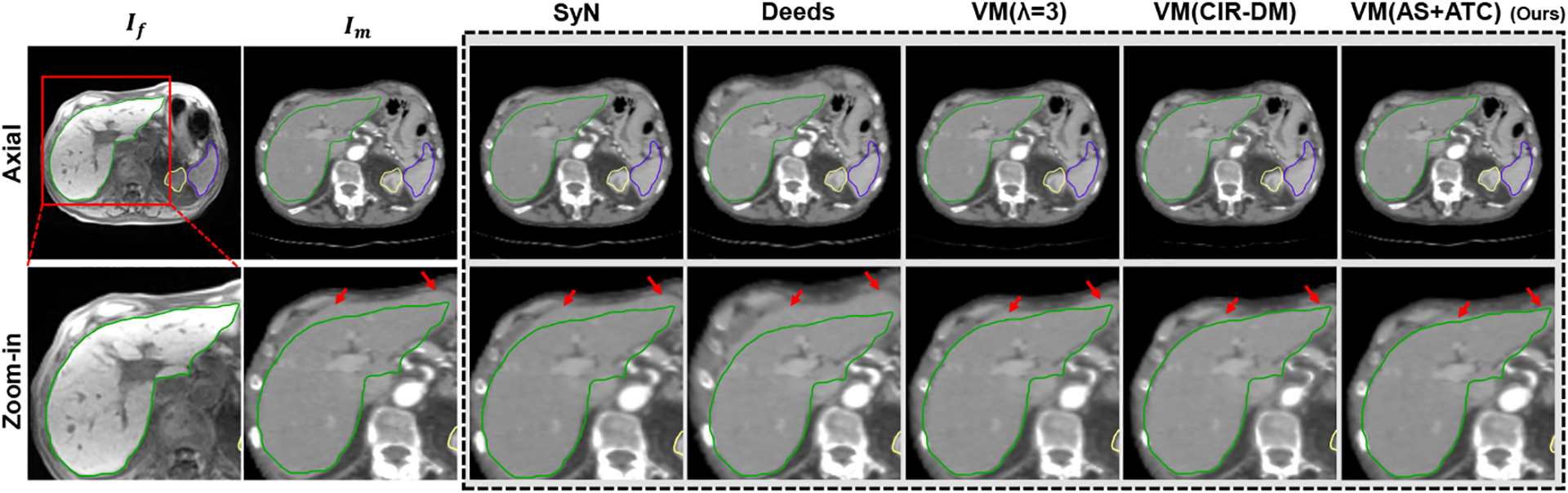
Exemplar axial slice of an abdominal CT-MRI registration case. The segmentation contours of the liver (green), kidney (yellow) and spleen (blue) extracted from the fixed abdominal MRI If are overlaid on all images. Better alignment drives structures closer to the fixed contours of If. The red arrows indicate the registration of interest around the organ boundary.