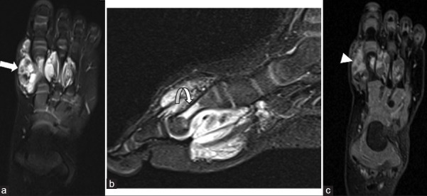
Figure 8.
Hemangioma (a) Axial T2 fat-saturated, (b) Sagittal STIR, and (c) Axial T1 fat-saturated post-contrast images shows an ill-defined infiltrating soft tissue mass lesion within the medial aspect of the forefoot. It demonstrates hyperintense signal on the T2 weighted images (arrows). Also note is made of a small hypointense foci within the mass on T2 weighted images which likely represents small phlebolith. On the post contrast images, there is heterogeneous enhancement (arrowhead). Note is made of intra-osseous extension into the 1st metatarsal shaft (curved arrow). STIR: Short tau inversion recovery

